Immunofluorescence patterns help eliminate “false positives” in diagnosing autoimmune rheumatic diseases
The detection of anti-nuclear antibodies, the ANA test, is a clear (laboratory-) diagnostic indicator of rheumatic autoimmune disease. One of the standard laboratory tests for the detection of these antinuclear antibodies is IIF, the indirect immunofluorescence assay, on human HEp-2 cells (ANA-HEp-2 test).

ANA-HEp-2 indirect immunofluorescence test (IIF): antibodies against RNP (ribonucleoproteins) – interphase nucleoli: coarse granular positive, nucleoli neglected; mitotic cells: negative (400x) – © ORGENTEC Diagnostika, Mainz
However, for up to 13% of healthy individuals, indirect immunofluorescence may detect anti-nuclear antibodies. Most of these healthy people will not develop an autoimmune disease – despite the positive ANA test. It is thus a challenge for the physician to differentiate these healthy, false-positive patients from those ANA-positive patients who already have an inflammatory rheumatic disease or who truly have an increased risk of developing such an autoimmune disease.
Several very specific IIF patterns
In a large study, Brazilian IIF experts have now worked out the fundamental differences between the ANA-HEp-2 test results on serum samples from healthy individuals and the immunofluorescence patterns from serum samples of patients with rheumatic disease; they have described various IIF patterns that can be used to differentiate between the two patient groups (Mariz et al. 2011). This study was published a few weeks ago in the January issue of Arthritis & Rheumatism, the journal of the American College of Rheumatology (ACR). In their article, the scientists from the Universidade Federal de São Paulo, Brazil, explain in detail that there are several very specific immunofluorescence patterns in the ANA-HEp-2 assay with which the autoimmune rheumatic diseases (ARD) are truly associated.
The investigations of Henrique A. Mariz, Emilia I. Sato, Silvia H. Barbosa, Silvia H. Rodrigues, Alessandra Dellavance, and Luis E. C. Andrade included 918 healthy people with ages ranging from 18 to 66 years. The results of the ANA-HEp-2 immunofluorescence tests (ANA-HEp-2 IIF assays) carried out on serum samples from these healthy participants were compared with those from a control group. The control group consisted of 153 patients who all suffered from an autoimmune rheumatic disease. They included 87 SLE patients (systemic lupus erythematodes), 45 patients with systemic sclerosis, 11 people with confirmed Sjögren’s syndrome, and 10 individuals suffering from idiopathic inflammatory myopathy (autoimmune myopathy).
Indirect immunofluorescence assays on ANA-HEp-2 cells were carried out on serum samples from all of these people. The result was evaluated as positive if the typical fluorescence pattern of indirect immunofluorescence was observed.
The results in brief: Well-defined IIF patterns, and thus a positive ANA-HEp-2 test result, were found for 12.9% of the healthy people. The ANA were observed at low to intermediate titres, and the nuclear fine speckled pattern (NFS) was particularly commonly observed – in nearly half of the ANA-positive healthy individuals (45.8%). The nuclear dense fine speckled pattern (NDFS) was found in nearly one third of the healthy people (33.1%), primarily in conjunction with a high ANA antibody titre. It is noteworthy that none of the healthy patients’ samples demonstrated the nuclear coarse speckled pattern (NCS) or a nuclear homogenous pattern (Ho).

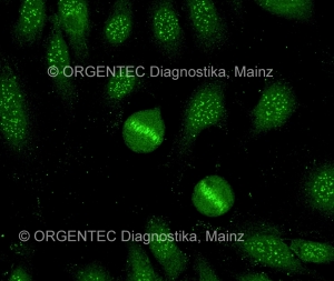
ANA-HEp-2 immunofluorescence pattern: antibodies against CENP-B (centromere) – interphase nuclei: the 46 dots are positive; mitotic cells are positive (400x) – © ORGENTEC Diagnostika, Mainz
In contrast, the serum samples from the autoimmune patients gave positive IIF results at intermediate to high ANA titres. The control group samples were also distinguishable by their completely typical fluorescence patterns. The serum samples from autoimmune patients were the only ones that demonstrated the nuclear coarse speckled (26% of samples), nuclear homogenous (7% of samples), nuclear centromeric (8%), and cytoplasmic dense fine speckled (3%) patterns. At 42%, the nuclear fine speckled pattern was also quite common in this group of patient serum samples; however, the antibodies were present at significantly higher titres than in the sample from healthy individuals. In a follow up after four years, 72.5% of the ANA-positive healthy people still had a positive ANA-HEp-2 test result. As before, they had no symptoms of autoimmune rheumatic disease!
ANA-HEp-2 pattern “is critical in properly diagnosing autoimmune disorders”
In a press release from Wiley-Blackwell (publisher of Arthritis & Rheumatism), Dr. Luis E. C. Andrade drew the following conclusion from his research group’s results: “The ANA-HEp-2 test is positive in a sizeable portion of the general population and our findings established distinguishing characteristics between healthy individuals and patients with autoimmune disease, which is essential to accurately interpret the test results.” In addition, “Our study confirms that the ANA-HEp-2 pattern is critical in properly diagnosing autoimmune disorders and future research should attempt to reproduce the interpretation of test results among different ANA experts and ANA-HEp-2 slide brands.”
[ … ] the ANA-HEp-2 pattern is critical in properly diagnosing autoimmune disorders and future research should attempt to reproduce the interpretation of test results among different ANA experts and ANA-HEp-2 slide brands.

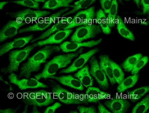
IIF pattern on the HEp-2 cells: antibodies against SRP (signal recognition particle) – cytoplasm: diffuse fine speckled positive; mitotic cells: negative (400x) – © ORGENTEC Diagnostika, Mainz
For many studies in autoimmune diagnostics and rheumatism diagnostics, the ANA test for HEp-2 cells is the method of choice; in fact, the ANA HEp-2 test system is often considered the gold standard of diagnosis. A few weeks ago, an Austrian colleague gave me basic instruction in the correct way to carry out indirect immunofluorescence tests and how to evaluate IIF patterns. I was amazed at how easy it is (in principle!) to carry out this technique. I was fascinated by the brilliance of the images I saw under the immunofluorescence microscope – and I was simultaneously humbled intimidated by the tremendous challenge involved in recognizing and evaluating the highly complex immunofluorecence patterns!
The analyst in an ordinary clinical laboratory should be able to recognize the ANA patterns that are clinically relevant and those that are the most probably observed in individuals with no apparent autoimmune disease.
As Dr. Andrade also points out in his article, careful and regular training, as well as a great deal of experience, are a requirement. In any case, he argues for basic knowledge of this highly complex topic: “The analyst in an ordinary clinical laboratory should be able to recognize the ANA patterns that are clinically relevant and those that are the most probably observed in individuals with no apparent autoimmune disease.” – I highly recommend you read the original text, it is worth it!
Re©ognise ©opyright law!!! — Please note that all immunofluorescence pictures used in this blog post are copyrighted material! All these IIF pattern fotos are taken and kindly left by Barbara Fabian, Austria. If you’re interested in using the pictures, please ask for permission: pr@orgentec.com
References:
Mariz HA, Sato EI, Barbosa SH, Rodrigues SH, Dellavance A, Andrade LE. Pattern on the antinuclear antibody-HEp-2 test is a critical parameter for discriminating antinuclear antibody-positive healthy individuals and patients with autoimmune rheumatic diseases. Arthritis Rheum. 2011 Jan;63(1):191-200. – doi: 10.1002/art.30084.
Author of this article: Tobias Stolzenberg

