The GRÜNER CLUB AUTOIMMUN blog featured a fine post about the history of indirect immunofluorescence. In that article my Austrian colleague Barbara Fabian, community manager of GRÜNER CLUB AUTOIMMUN, described in great detail how indirect immunofluorescence technology, or: IFT, and also referred to as IIF assay, has become an indispensible tool of autoimmune disease diagnostics over the last two decades, and how IFT has become a standard laboratory technique used in serological autoimmune diagnostics.
Without further ado I have translated Barbara’s post in order to make you this text, and especially the interesting images, available. – Here it is:
The Development of Indirect Immunofluorescence Technology (IFT)
by Barbara Fabian, MSc, Community Manager of GRÜNER CLUB AUTOIMMUN
Over the last 20 years, the detection of autoantibodies has developed into an indispensible component of autoimmune diagnostics. Along with serological and clinical data, autoimmune status has become an important building block in the formation of diagnoses.
Autoantibodies were first detected many years ago on frozen sections of mouse and rat liver. The samples were generally prepared in the laboratory, and the other required reagents, such as conjugates, were rarely available at the optimal concentrations required for the individual diagnostic test systems. Early autoimmune results were thus found to be highly variable. The use of different substrates for autoantibody detection showed that autoantibodies against nuclear components, such as those against double-stranded DNA, anti-dsDNA antibodies, which exhibited a homogeneous immunofluorescence pattern, were easily detectable on liver sections (Figure 1).

Figure 1: Indirect immunofluorescence test (IFT) on tissue sections: homogeneous immunofluorescence pattern on mouse kidney and stomach – © Barbara Fabian, www.der-gruene-club.at
The HEp-2 cell line raises new possibilities
However, not all antinuclear antibodies (ANA) exhibit a typical immunofluorescence pattern on mouse or rat organ tissue sections. For this reason, the additional detection of autoantibodies on human epithelial cells, known as HEp-2 cells, was developed after a few years. The use of this cell line has the advantage that a very broad spectrum of autoantibodies is detectable on HEp-2 cells. Antibodies against cell-cycle dependent antigens exhibit no immunofluorescence pattern on organ tissue sections. However, they are, like proliferating cell nuclear antigen (PCNA), of significance in the diagnosis of systemic lupus erythematosus (SLE), and can be detected on HEp-2 cells in the autoimmune laboratory (Figure 2).
Because both the mitotic phase and metaphase are identifiable in these cells, information regarding the patterns of the chromosomes is also available (Bayer PM, Fabian B, Hübl W: Immunofluorescence Assays (IFA) and Enzyme-Linked Immunosorbent Assays (ELISA) in Autoimmune Disease Diagnostics – Technique, Benefits, Limitations and Applications. – The link leads to the PubMed abstract). This allowed new, previously undetectable antibodies against the HEp-2 cell cytoplasm (cytoplasmic antibodies), such as the anti-SRP antibodies (SRP stands for signal recognition particle) or antibodies against PL7 or PL12 (anti-PL7, anti-PL12), to be detected as well. This led to numerous studies regarding the clinical significance of these new biomarkers. Various diagnostics companies that specialized in the development of test systems for the detection of autoimmune diseases developed tests for these “new” autoantibodies, such as the cytoplasm immunoblot test system.

Figure 2: IFT in autoimmune disease diagnostics: laboratory detection of anti-PCNA antibodies on HEp-2 cells – © Barbara Fabian, www.der-gruene-club.at
In order to establish the value of the HEp-2 cell in the diagnosis of antinuclear antibodies, these cells were initially compared to the liver. It was demonstrated that the sensitivity of antinuclear antibodies, ANA, in human cells was better than that in tissue sections. Rat kidney and rat liver demonstrated the least sensitivity in the detection of nuclear antibodies (Hahon N, Eckert HL, Stewart J: Evaluation of Cellular Substrates for Antinuclear Antibody Determinations. – The link leads to a free full-text article in the Journal of Clinical Microbiology).
To this day, antibody detection on HEp-2 cells has retained its central role. When autoimmune disease is suspected, the HEp-2 test is used for screening, which allows for the cost-effective and high-quality serological diagnosis of various autoimmune diseases (Hiemann R et al. Challenges of Automated Screening and Differentiation of Non-Organ-Specific Autoantibodies on HEp-2 Cells. – this link leads to an abstract in Autoimmunity Reviews). The sensitive detection of many clinically relevant autoantibodies is possible on HEp-2 cells. Any positive result is followed up by a step-wise diagnosis in which, depending on the evaluation of the reactivity with HEp-2 cells, other substrates may be used or other immunological tests like ELISA (enzyme-linked immunosorbent assay) or immunoblot assays carried out.
Tissue sections – indispensible for gastroenterological diagnostics
Diagnosis by means of tissue sections remains very important in gastroenterology. In cases of autoimmune hepatitis (AIH) especially, the detection of anti-smooth muscle antibodies, or: ASMA, or antibodies to liver-kidney microsomes, or: anti-LKM antibodies, are important diagnostic tools. Testing on HEp-2 cells alone would not suffice in these cases, because tests for anti-smooth muscle antibodies, the so called ASMA tests, are not always positive, and tests for anti-LKM autoantibodies are not positive. If autoimmune hepatitis is suspected, routine diagnostics call for testing for antibodies on mouse or rat tissues. If required, a positive antibody screening test can be confirmed by enzyme immunoassays (“ELISA tests”) or immunoblot tests (“blot assays”).
Autoantibodies may be detectable years before the diagnosis of an autoimmune disease. For example, the antimitochondrial antibodies (AMA) in cases of primary biliary cirrhosis (PBC), which are also detected by means of mouse or rat tissue (Beleznay Z, Regenass S. Diagnostics of Autoimmune Diseases. – The link leads to an English language abstract in PubMed, the article itself is in German). A consensus report published in 2004 included guidelines and recommendations in this regard (Vergani D et al. Liver Autoimmune Serology: a Consensus Statement from the Committee for Autoimmune Serology of the International Autoimmune Hepatitis Group. – This link leads to a free full-text article in the Journal of Hepatology). The report recommends diagnosis on a section consisting of combined kidney, stomach, and liver tissue (KSL). It also made the first steps toward standardization by establishing the optimal size and histological composition of the tissues. First attempts at analytical standardization, including incubation times, for example, were also covered in this publication.
Indirect immunofluorescence tests (IFT assays) in ANCA diagnostics
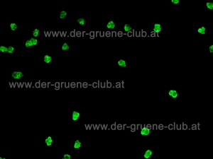
Another important area in the diagnosis of autoimmune diseases is the serological diagnosis of systemic autoimmune vasculitis, such as Wegner’s granulomatosis or microscopic polyangiitis. This is achieved through the detection of autoantibodies against cytoplasmic neutrophil granulocytes with the ANCA test (ANCA = anti-nuclear cytoplasmic antibodies). ANCA primarily involves antibodies that display perinuclear (pANCA) or cytoplasmic (cANCA) staining upon exposure to ethanol-fixed granulocytes (Figure 3). In addition to ethanol-fixed granulocytes, formalin- and methanol-fixed granulocytes are also used.
The additional use of formalin-fixed granulocytes allows for the differentiation of the antinuclear antibodies from the ANCA test. Methanol-fixed neutrophil granulocytes are rarely used in routine laboratory analysis because they deliver relatively little information. Diagnostic tests like ELISA and immunoblot assays that are directed toward myeloperoxidase (anti-MPO test systems) and proteinase 3 (anti-PR3 assays) are also available for autoantibodies against neutrophil granulocytes.
A consensus study on the subject of ANCA diagnostics was published in 1999, with an addendum issued in 2003 (Savige J et al. International Consensus Statement on Testing and Reporting of Antineutrophil Cytoplasmic Antibodies (ANCA). – This link leads to the PubMed abstract. – Savige J. et al. Addendum to the International Consensus Statement on Testing and Reporting of Antineutrophil Cytoplasmic Antibodies. Quality Control Guidelines, Comments, and Recommendations for Testing in other Autoimmune Diseases. – This link leads to the full-text article in the American Journal of Clinical Pathology.)
The emphasis of these studies is the clinical importance of tests for antibodies against neutrophil granulocytes. However, the goals of these studies also included the standardization of indirect immunofluorescence techniques for the detection of ANCA, including recommendations for the optimal dilution of the serum samples, the conjugate, and the counterstain. A diagnostic scheme for both minimal and optimal antibody detection was established.

Figure 3a: IFT detection of pANCA on ethanol-fixed neutrophil granulocytes, ANCA diagnosis with the aid of indirect immunofluorescence technology – © Barbara Fabian, www.der-gruene-club.at
The continuing issue of standardization
Over the years, many publications have discussed the analysis of autoantibodies through indirect immunofluorescence. The emphasis of a 1983 study on the problems with the standardization of immunofluorescence was on the reproducibility of the detection of antinuclear antibodies, known as ANA detection (Beutner EH, Krasny S, Kumar V, Taylor R, Chorzelski TP. Prospects and Problems in the Definition and Standardization of Immunofluorescence. Present Levels of Reproducibility and Disease Specificity of Antinuclear Antibody Tests. – This link leads directly to the article in the Annals of the New York Academy of Sciences). In addition to pointing out the fact that certain autoantibodies such an anti-centromere antibodies react with human cell lines while remaining undetectable on tissue sections or mouse fibroblast cells, this work also covers methodological studies regarding the titre of antibodies.

Figure 3b: IFT detection of pANCA on formalin-fixed neutrophil granulocytes. – © Barbara Fabian, www.der-gruene-club.at
In order to minimize variations in titre between different laboratories, the influence of the conjugate was described. At the center of the investigation were the specificity of the concentration, the dilution, and the fluorescein/protein ratio of the conjugate used. The sensitivity of the optical systems used to evaluate the sample slides was also compared. The use of optically standardized sample slides was recommended as a means to control the variation between different systems.
The quality of test products has improved sharply over time. Because diagnostics firms now offer prepared test kits for the serodiagnosis of autoimmune diseases, which contain mutually calibrated “ready-to-use” reagents, the technical aspect of work in the field of autoimmune diagnostics has been significantly simplified.
However, diagnosis by means of indirect immunofluorescence continues to demand a great deal of knowledge and experience, continuing to offer a challenge for laboratory personnel and physicians. This challenge is surely also the reason why most people working in this field exhibit great enthusiasm and intensity in tackling this problem, and why advanced training in this field is of great value.
Please note: All fotographs of immunofluorescence patterns in this article are protected by copyright law, which is ensured by watermarks. If the pictures have attracted your interest, and if you feel the interest to use one or a number of immunofluorescence pictures for a lecture, a presentation, a publication … please feel free to get into contact with Barbara Fabian via the GRÜNER CLUB AUTOIMMUN website.
Author of this article: Tobias Stolzenberg
References:
Bayer PM, Fabian B, Hübl W. Immunofluorescence assays (IFA) and enzyme-linked immunosorbent assays (ELISA) in autoimmune disease diagnostics–technique, benefits, limitations and applications. Scand. J. Clin. Lab. Invest. Suppl. 235, 68–76 (2001). – link leads to the abstract in PubMed: http://www.ncbi.nlm.nih.gov/pubmed?term=Bayer%20Fabian%20H%C3%BCbl
Beleznay Z, Regenass S. [Diagnostics of autoimmune diseases]. Ther Umsch 65, 529–537, doi:10.1024/0040-5930.65.9.529 (2008). – link leads to the English-language abstract in PubMed, the article itself is written in German: http://www.ncbi.nlm.nih.gov/pubmed?term=Beleznay%20Regenass
Beutner EH, Krasny S, Kumar V, Taylor R, Chorzelski TP. Prospects and problems in the definition and standardization of immunofluorescence. I. Present levels of reproducibility and disease specificity of antinuclear antibody tests. Ann. N. Y. Acad. Sci. 420, 28–54 (1983). – link leads to the full-text article in the Annals of the New York Academy of Sciences: http://onlinelibrary.wiley.com/doi/10.1111/j.1749-6632.1983.tb22186.x/abstract;jsessionid=1AC6AD5E6E4751591D0CE810B3E1E6D7.d02t02
Hahon N, Eckert HL, Stewart J. Evaluation of cellular substrates for antinuclear antibody determinations. J. Clin. Microbiol. 2, 42–45 (1975). – link to the free full-text article in the Journal of Clinical Microbiology: http://jcm.asm.org/cgi/reprint/2/1/42?view=long&pmid=818105
Hiemann R et al. Challenges of automated screening and differentiation of non-organ specific autoantibodies on HEp-2 cells. Autoimmun Rev 9, 17–22, doi:10.1016/j.autrev.2009.02.033 (2009). – link leads to the abstract in Autoimmunity Reviews: http://www.sciencedirect.com/science/article/pii/S1568997209000731
Savige J et al. International Consensus Statement on Testing and Reporting of Antineutrophil Cytoplasmic Antibodies (ANCA). Am. J. Clin. Pathol. 111, 507–513 (1999). – link leads to the PubMed abstract: http://www.ncbi.nlm.nih.gov/pubmed/10191771
Savige J. et al. Addendum to the International Consensus Statement on testing and reporting of antineutrophil cytoplasmic antibodies. Quality control guidelines, comments, and recommendations for testing in other autoimmune diseases. Am. J. Clin. Pathol. 120, 312–318, doi:10.1309/WAEP-ADW0-K4LP-UHFN (2003). – link leads to the full-text article in the American Journal of Clinical Pathology: http://ajcp.ascpjournals.org/content/120/3/312.long


2 Comments
Add a Comment